BodyWorks Eve

Realistic, safe & effective training in ultrasound-guided needling

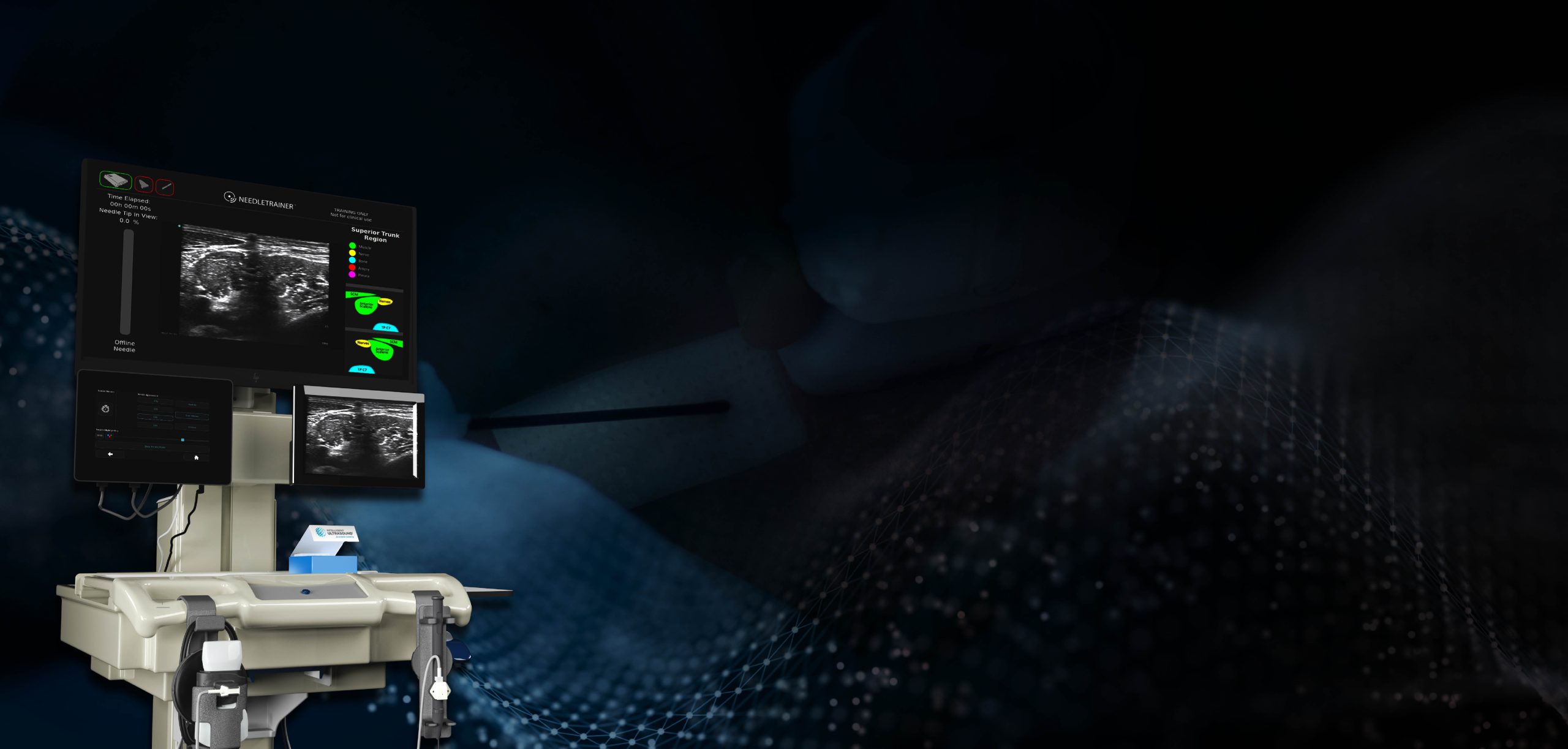
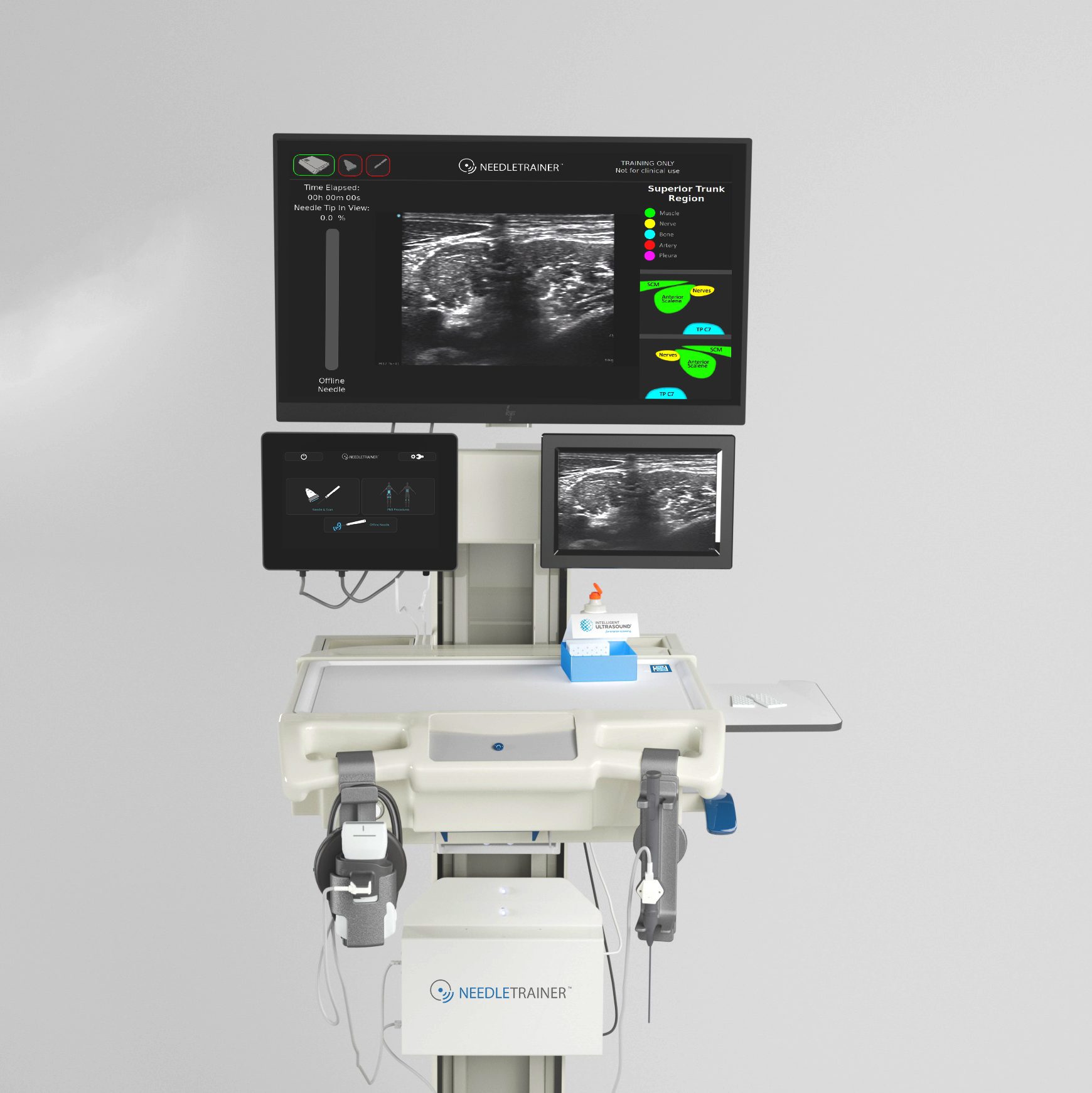
NeedleTrainer™ provides an all-in-one training solution to teach ultrasound-guided needling safely and effectively, using non-invasive needle insertion during real-time scanning on a real subject.
Needle Trainer

Now available to even more institutions

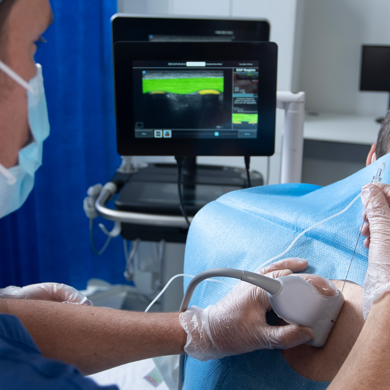
The first of its kind, NeedleTrainer™ uses a retractable needle and virtual image overlays to simulate needling non-invasively on a live participant, using an authentic live ultrasound scan. This enables trainees to develop hand-eye coordination, optimum positioning, and accuracy in ultrasound-guided interventional procedures in a realistic clinical environment with minimal risk.

NeedleTrainer™ is the latest development in Intelligent Ultrasound’s vision to make ultrasound simpler to use and easier to learn. The new generation of NeedleTrainer™ encompasses a GE Healthcare Vscan™ Air providing an all-in-one training solution for any medical education program teaching ultrasound-guided needling, enabling centers to adapt to new curriculum requirements on learning these skills without impacting clinical hours or patient safety.
Needle Trainer

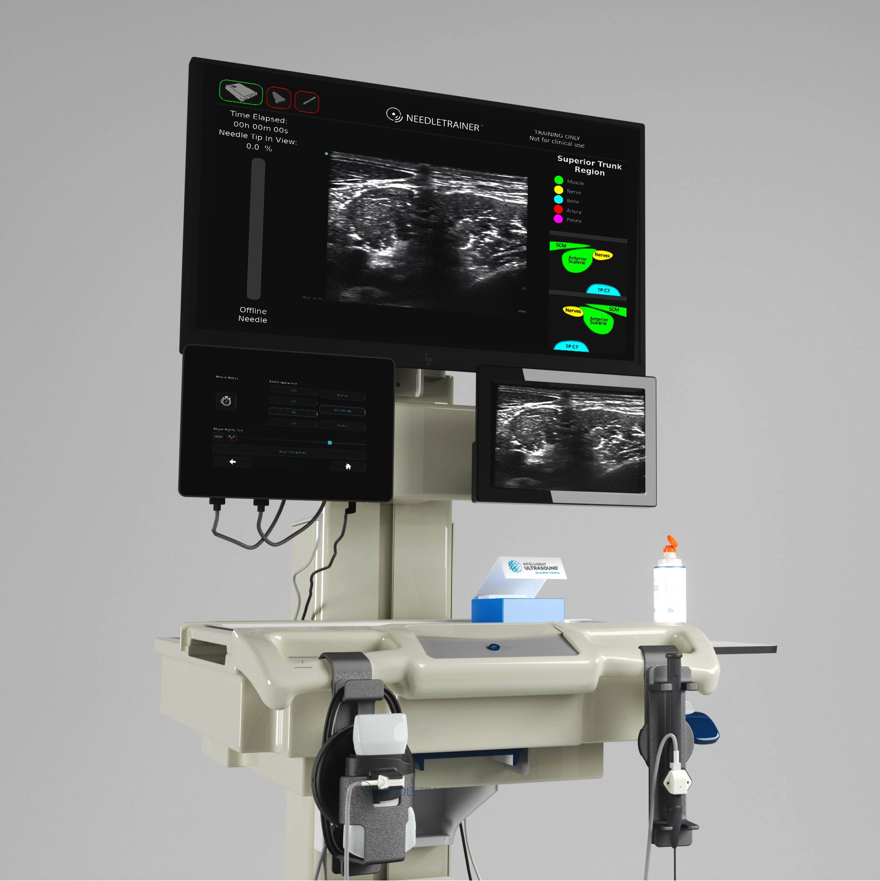
Metrics for proficiency of needle probe co-ordination

Needle visualization measurements provide objective quantitative metrics for needle probe alignment, to aid assessment in proficiency and competence of needle probe coordination:

Time elapsed (s)
Tip in view (%)

Needle Trainer

Tailor needle specifications to your procedure

Allows customization of the virtual needle simulation according to the procedure & specialty requirements:

Echogenicity: Obvious, Semi-obvious, Realistic
Gauge: 14G, 18G, 22G and 27G

Needle Trainer

Complete learning pathway for regional anesthesia

Extend learning to include image interpretation along with needle-probe coordination, with the full classroom-to-clinic learning package*, specifically designed for regional anesthesia.

*The classroom-to-clinic package includes NeedleTrainer plus, with anatomy highlighting of 10 peripheral nerve blocks, and ScanNav Anatomy PNB.
PNB

ScanNav Anatomy Peripheral Nerve Block (PNB)

ScanNav Anatomy PNB is designed to enhance the accuracy and standardisation of ultrasound image interpretation by providing a real-time colour overlay of key sono-anatomical structures.

Enabling more qualified healthcare professionals to perform ultrasound-guided regional anaesthesia with safety and accuracy

INFORMATION ABOUT US

CONTACT US FOR ANY QUESTIONS